Thomas Shane, M.D.
Contributing Editor
Case History:
A 20 year old male presents to the oculoplastics service with a chief complaint of a right brow mass. The patient states that the mass is painless and has been slowly enlarging over the previous two years. He denies any past ocular history or trauma.
Past medical history is positive for tonsillectomy. The patient takes no medicines. Family and social history are non-contributory. Review of systems is negative.
On clinical examination, the patient is alert, and oriented. His visual acuity is 20/25 in both eyes. Examination of the right brow reveals a soft, non-tender mass approximately 2 cm x 1 cm in dimension (Figure 1). The mass is pink and pulsatile in nature, with an audible bruit upon auscultation. There is no globe displacement or proptosis. The remainder of the ocular exam is within normal limits.
Ultrasound of the mass shows an irregularly shaped network of tubular, low-reflective, high-flow lesions filling the anterior superotemporal orbit (Figure 2). The lesions do not enlarge on valsalva. MRI of the mass demonstrates prominent vascular flow voids (Figure 3).
Diagnosis: Orbital Arteriovenous Malformation (AVM)
Orbital arteriovenous malformations are rare, responsible for only 0.5% of all orbital tumors. AVM’s are vascular lesions consisting of arteries and veins that directly communicate in the absence of a capillary bed. This communication creates a high-flow nidus of vessels that slowly enlarges with time, changes in hormonal states, or trauma.
Clinically, orbital AVM’s are most distinguishable by their pulsatile nature. They are one of the few diagnoses in ophthalmology that require the use of a stethoscope (a refreshing reminder of our previous medical education). Audible bruits can be heard and sometimes a palpable thrill can be felt over these unique lesions.
Imaging of these lesions can be accomplished with any of the following modalities: ultrasound, CT, or MRI. But the gold standard for imaging these vascular malformations is conventional angiography (Figure 4). This technique allows for direct examination of the feeding arteries, nidus, and draining veins of the AVM in preparation for surgical management.
Treatment of AVM’s is conservative, given that the majority of these lesions are asymptomatic. The potential for hemorrhage or mass effect still exists, however, in which case aggressive treatment becomes necessary. Surgical excision has been used with success in the past, but with risk of exsanguination or collateral damage to delicate neighboring structures.
One alternative to surgical excision is endovascular embolization, which has grown by leaps and bounds in recent years. Thanks to the advent of microcatheters, previously unreachable vessels of extremely small caliber can now be cannulated. Perhaps the most exciting development in endovascular embolization is the advent of several new embolic agents, each with unique characteristics:
- Polyvinyl alcohol is administered in fine particles that are thrombogenic when
infused into small arteries, markedly reducing flow through the AVM.
- n-Butyl cyanoacrylate is an adhesive monomeric liquid that quickly polymerizes and becomes solid when exposed to blood. This leads to occlusion and permanent fibrosis of the AVM feeder vessels.
- Ethylene-vinyl alcohol copolymer, a non-adhesive solution, allows for longer and more controlled infusions without limit of polymerization.
Using these agents, AVM’s can be markedly reduced in preparation for definitive excision or obliterated altogether. Indeed, the success of endovascular embolization has made it a very popular treatment in today’s management of AVM’s. (Figure 5).
Given the superficial and asymptomatic nature of his AVM, our patient is currently being managed with conservative therapy. Should the AVM become painful, hemorrhagic, a threat to vision, or cosmetically unacceptable, embolization followed by surgical excision will be the likely course of action.
Figure 1: Pink, pulsatile right lateral brow mass.
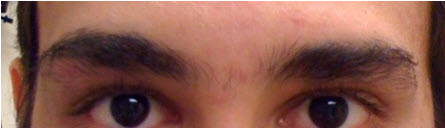
Figure 2: Ultrasound reveals an irregularly shaped network of tubular, low-reflective lesions with marked blood flow filling the anterior superotemporal orbit

Figure 3: MRI showing prominent vascular flow voids

Figure 4: Cerebral angiogram demonstrating multiple feeding arteries (small arrows) and central nidus (large, grey arrow) characteristic of an arteriovenous malformation.

Figure 5: Cerebral angiogram of an AVM following embolization with n-butyl cyanoacrylate. Notice the lack of feeder vessels and blood flow to the nidus of the AVM.

- Hayes BH, et al. “Management of Orbital and Periorbital Arteriovenous Malformations.” Ophthalmic Surgery Mar/Apr, 1995, 26(2): 145-152.
- Decock, CD, et al. “Diagnosis and Treatment of a Superficial Upper Eyelid Arteriovenous Malformation.” Orbit. 2008, 27:301-3.
- Linfante I, Wakhloo AK. “Brain Aneurisms and Arteriovenous Malformations: Advancements and Emerging Treatments in Endovascular Embolization.” Stroke. April 2007. 38(4): 1411-7.
Have a question or comment on this article? Use the "Comment" link above to leave your thoughts, and the author will respond.